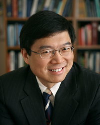
 Lihong Wang earned his Ph.D. degree at Rice University, Houston, Texas under the tutelage of Robert Curl, Richard Smalley, and Frank Tittel. He currently holds the Gene K. Beare Distinguished Professorship of Biomedical Engineering at Washington University in St. Louis. His book entitled “Biomedical Optics: Principles and Imaging,” one of the first textbooks in the field, won the 2010 Joseph W. Goodman Book Writing Award. He also edited the first book on photoacoustic tomography and coauthored a book on polarization. He has published 437 peer-reviewed articles in journals including Nature (Cover story), Science, PNAS, and PRL and delivered 433 keynote, plenary, or invited talks. His Google Scholar h-index and citations have reached 99 and 38,700, respectively. His laboratory was the first to report functional photoacoustic tomography, 3D photoacoustic microscopy (PAM), photoacoustic endoscopy, photoacoustic reporter gene imaging, the photoacoustic Doppler effect, the universal photoacoustic reconstruction algorithm, microwave-induced thermoacoustic tomography, ultrasound-modulated optical tomography, time-reversed ultrasonically encoded (TRUE) optical focusing, nonlinear photoacoustic wavefront shaping (PAWS), compressed ultrafast photography (100 billion frames/s), Mueller-matrix optical coherence tomography, and optical coherence computed tomography. In particular, PAM broke through the long-standing diffusion limit on the penetration of optical microscopy and reached super-depths for noninvasive biochemical, functional, and molecular imaging in living tissue at high resolution. He has received 37 research grants as the principal investigator with a cumulative budget of over $47M. He is a Fellow of the AIMBE (American Institute for Medical and Biological Engineering), Electromagnetics Academy, IEEE (Institute of Electrical and Electronics Engineers), OSA (Optical Society of America), and SPIE (Society of Photo-Optical Instrumentation Engineers). He is the Editor-in-Chief of the Journal of Biomedical Optics. He chairs the annual conference on Photons plus Ultrasound. He was a chartered member of an NIH Study Section. Wang serves as the founding chairs of the scientific advisory boards of two companies, which have commercialized photoacoustics. He received NIH’s FIRST, NSF’s CAREER, NIH Director’s Pioneer, and NIH Director’s Transformative Research awards. He also received the OSA C.E.K. Mees Medal, IEEE Technical Achievement Award, IEEE Biomedical Engineering Award, SPIE Britton Chance Biomedical Optics Award, and Senior Prize of the International Photoacoustic and Photothermal Association for “seminal contributions to photoacoustic tomography and Monte Carlo modeling of photon transport in biological tissues.” An honorary doctorate was conferred on him by Lund University, Sweden. His lab is transitioning to Caltech but plans to continue his collaborations with the medical school of Washington University
Redefining the Spatiotemporal Limits of Optical Imaging: Photoacoustic Tomography, Wavefront Engineering, and Compressed Ultrafast Photography
Abstract: Photoacoustic tomography has been developed for in vivo functional, metabolic, molecular, and histologic imaging by physically combining optical and ultrasonic waves. Broad applications include early-cancer detection and brain imaging. Conventional high-resolution optical imaging of scattering tissue is limited to depths within the optical diffusion limit (~1 mm in the skin). Taking advantage of the fact that ultrasonic scattering is orders of magnitude weaker than optical scattering per unit path length, photoacoustic tomography beats this limit and provides deep penetration at high ultrasonic resolution and high optical contrast. The annual conference on photoacoustic tomography has become the largest in SPIE’s 20,000-attendee Photonics West since 2010.
Wavefront engineering overcomes the optical diffusion limit by actively controlling the wavefront of the incident light to optimize the light intensity at a target position inside tissue. One such technology is called time-reversed ultrasonically encoded (TRUE) optical focusing, which can noninvasively deliver light to a dynamically defined focus deep in a scattering medium. First, diffused coherent light is encoded by a focused ultrasonic wave to provide a virtual internal “guide star”; then, only the encoded light is time-reversed and transmitted back to the ultrasonic focus.
Compressed ultrafast photography (CUP) can image in 2D non-repetitive time-evolving events at up to 100 billion frames per second. CUP has a prominent advantage of measuring an x, y, t (x, y, spatial coordinates; t, time) scene with a single camera snapshot, thereby allowing observation of transient events occurring on a time scale down to tens of picoseconds. Further, akin to traditional photography, CUP is receive-only—avoiding specialized active illumination required by other single-shot ultrafast imagers. Given CUP’s capability, we expect it to find widespread applications in both fundamental and applied sciences including biomedical research.

